Assessing Complementary Information on Stroke Effects
PET and MRI have been used to study stroke in small animal models, examining changes in water diffusion and glucose metabolism. Ischemic events produce a cascade of rapidly evolving physiological changes[1]The science of cerebral ischemia and the quest for neuroprotection: navigating past failure to future success, Turner et al. J Neurosurg. 2013, including a reduction in cerebral blood flow (CBF) and cerebral blood volume (CBV), that can be highly individualized in certain animal models, creating data interpretation challenges when animals are imaged sequentially. With instruments capable of simultaneous PET/MRI acquisition researchers can assess complimentary information about the evolution of injury and the effect of interventions such as neuroprotective compounds.
MRI has been used extensively to monitor water diffusion changes in stroke patients, quantified by the apparent diffusion coefficient (ADC) using diffusion weighted imaging (DWI). PET provides complimentary information, and has been used to evaluate changes in glucose metabolism in rat models.
Benefits of Simultaneous PET/MRI to Observe Heterogenous Changes During Brain Injury
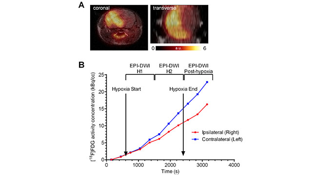
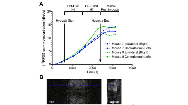
Recently a group of researchers from the University of California, Davis and Genentech demonstrated successful multi-modal evaluation of the time course of a hypoxic challenge in mouse models of cerebral hypoxia-ischemia[2]Simultaneous PET/MRI Imaging During Mouse Cerebral Hypoxia-ischemia, Yu Ouyang et al. J. Vis. Exp, 2015.. The extent of brain injury is variable in these models[3]Strain-related brain injury in neonatal mice subjected to hypoxia–ischemia, R. Ann Sheldon et al. Brain Research, 1998., and along with the speed of ischemic changes, make a strong argument for simultaneous PET/MRI acquisition in small-animal stroke research.
In most animals, [18F]FDG PET images fused with anatomical MRI images showed reduced uptake in the occluded side relative to the non-occluded side, although this was not true in all cases likely due to animal variability. Executing sequential imaging studies and relying on software co-registration techniques would require the assumption that animal physiology has not significantly changed between imaging sessions; a potential pitfall given the rapid progression of ischemic brain injury.[4]Simultaneous PET/MRI Imaging During Mouse Cerebral Hypoxia-ischemia, Yu Ouyang et al. J. Vis. Exp, 2015. Simultaneous PET/MRI instrumentation can arm researchers with a new, powerful tool for multi-modal investigation into physiological changes during stroke and the effectiveness of novel interventional strategies.

References
| ↑1 | The science of cerebral ischemia and the quest for neuroprotection: navigating past failure to future success, Turner et al. J Neurosurg. 2013 |
|---|---|
| ↑2, ↑4 | Simultaneous PET/MRI Imaging During Mouse Cerebral Hypoxia-ischemia, Yu Ouyang et al. J. Vis. Exp, 2015. |
| ↑3 | Strain-related brain injury in neonatal mice subjected to hypoxia–ischemia, R. Ann Sheldon et al. Brain Research, 1998. |