Correlate Brain Chemistry with Brain Activity
Both PET and MRI scanning have been used for neuroimaging[1]Behind the scenes of functional brain imaging: A historical and physiological perspective, Marcus E. Raichle, Proc. Natl. Acad. Sci. USA, Vol. 95, pp. 765-772, Feb 1998. However, with the advent of simultaneous PET/MR imaging, these complementary techniques allow researchers to better correlate changes in brain chemistry with changes in brain activity.
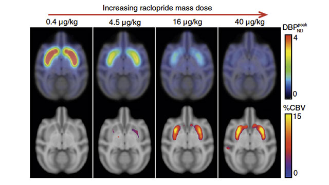
Simultaneous PET/fMRI Measures Response to a Pharmaceutical Antagonist
A recent study by researchers from Massachusetts General Hospital, MIT, the University of Copenhagen, and Katholieke Universiteit in Leuven employed simultaneous PET/fMRI to measure neurovascular response when administering pharmacologic doses of a receptor agonist while at the same time measuring dopamine D2/D3R occupancy in basal ganglia of non-human primates.[2]Neurovascular coupling to D2/D3 dopamine receptor occupancy using simultaneous PET/functional MRI, Christin Y. Sander, et. al., Proc. Natl. Acad. Sci. USA, Vol. 110, No. 27, pp. 11169–11174, Jul … Continue reading.
The results showed strong correlation between neurovascular response and dopamine receptor binding across a wide dynamic range and demonstrate the usefulness of concurrent assessment of hemodynamics and receptor-specific neurotransmission in preclinical and clinical studies.

Recent Development of PET/MRI Capability Enables New Research
Prior to this study, little or no reporting had been done on comparing the functional output measured by fMRI and changes to the neuroreceptor system target by a specific ligand. The authors suppose that this absence of published work is due to the lack of suitable instrumentation such as a combined PET/fMRI system for simultaneous quantitative imaging. PET emission data offers high sensitivity and neurochemical specificity, while fMRI offers high spatio-temporal resolution for changes in neurovascular activity.
In a few short years, simultaneous PET/MRI has become more commonplace. As of 2015, the Whole Brain Atlas[3]The Whole Brain Atlas, a collaboration between Harvard University and MIT and hosted online by the Harvard University Medical School, can display high-resolution MRI, PET, as well as combined PET/MRI … Continue reading even gives the viewer the ability to visualize both PET and MRI images of normal anatomy separately as well as together.

References
| ↑1 | Behind the scenes of functional brain imaging: A historical and physiological perspective, Marcus E. Raichle, Proc. Natl. Acad. Sci. USA, Vol. 95, pp. 765-772, Feb 1998 |
|---|---|
| ↑2 | Neurovascular coupling to D2/D3 dopamine receptor occupancy using simultaneous PET/functional MRI, Christin Y. Sander, et. al., Proc. Natl. Acad. Sci. USA, Vol. 110, No. 27, pp. 11169–11174, Jul 2013 |
| ↑3 | The Whole Brain Atlas, a collaboration between Harvard University and MIT and hosted online by the Harvard University Medical School, can display high-resolution MRI, PET, as well as combined PET/MRI images. |